
Featured Research
All kinds of important and interesting research is being performed on Aquaneering systems all over the world. Click here to view a variety of articles detailing experiments that use our systems. Below, you can find a variety of case studies showcasing what we do to help our customers.

Slater’s initial eighth grade research featured these zebrafish in an Aquaneering eRack.
In 2017, the University of Arizona awarded Slater with a scholarship.

In 2019, Slater returned to Dr. Welsh’s classroom to fix an eRack pump.
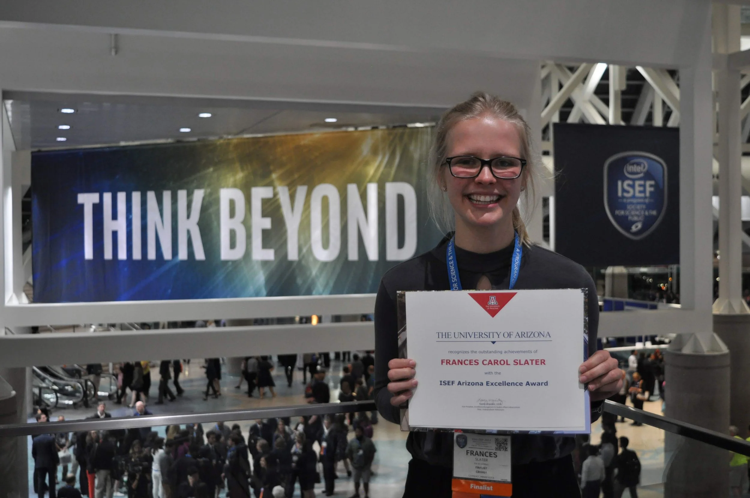
Frances Slater, climate scientist and activist
In 2013, Dr. Cynthia Welsh, a seventh-grade life science teacher/science research teacher from Cloquet, MN, and Dr. Jennifer Liang, University of Minnesota Duluth Biology professor, collaboratively wrote a proposal to obtain an eRack from AQUANEERING Inc. The eRack was obtained and setup in Dr. Welsh’s seventh grade classroom. Since then, over 2,000 seventh graders have learned about genetics and taking care of zebrafish in their classroom. Many seventh graders did independent research projects using the system. Many of these students continued to use the eRack in high school for more complicated research, all of them participating and winning awards in regional, state, and international science fairs.
One of those students, Frances Slater, has a research story that illustrates how having an eRack available in the classroom for research lead her down a path to win regional, state, and international science fair awards. Eventually, it also led to a professional publication of her work and a full tuition scholarship to the University of Arizona.
In eighth grade, Slater approached her classroom mentor, Dr. Welsh, and they discussed how she wanted to focus her research project on the study of genetics. Dr. Welsh told her that genetics might be a difficult topic to tackle in eighth grade yet she would try and find someone to help her. One of her then-high school students, Anna Pollak, was doing genetic zebrafish work with her professional mentor, Dr. Liang, who agreed to help. These projects and collaborations are what lead to Dr. Welsh and Liang writing the grant proposal to obtain an eRack.
As an eighth grader with a desire to study genetics, Slater decided a simple Punnett Square and Chi Square analysis of mating pairs of transgenic fluorescent zebrafish would be an interesting place to start. Slater set up the eRack and conditioned it on her own. She monitored tank chemistry until she knew it was ready for the study zebrafish to arrive. Using the eRack, Slater compared the outcome of her mating pairs to what one would expect to see by phenotype (color). This research was titled: One Fish, Two Fish, Red Fish, Blue Fish: What effect does the genotype of a zebrafish cross have on the phenotype of their offspring? For this work, she advanced from the NE MN Regional Science Fair to the MN Academy of Science State Science Fair. At the state fair, she received the Gold Medal award, the top Grand Award at the fair. Slater also received the Seagate Emerging Scientist Award, which recognizes excellence in scientific research conducted by middle school students, and the Broadcom Masters Award given to the top 10 percent of middle school projects.
Using the eRack, Slater continued to do increasingly more complex science projects. As a freshman in high school, she broadened her research focus to evaluate the effect of the inserted transgene on zebrafish fitness. Her paper was titled: Transgenic Fluorescent Zebrafish: The use of growth and dissolved oxygen consumption per mass of fish to determine the impact of gender, transgenes, and zygosity on the fitness and impact in the wild. She presented this research at the NE MN Regional Science Fair and won numerous awards including the Zookeeper Award, MN Chapter of American Fisheries Award, first place research paper award, advancing her to present at the Junior Science and Humanities Symposium (JSHS), and first place project award, advancing her to the Intel International Science and Engineering Fair (Intel ISEF) in Pittsburgh, PA. At the State science fair, her poster presentation was equally successful, again winning the American Fisheries Society Aquatic Science Award of Excellence, as well as the US Metric Association Award and the Wolfram Research Mathematica Software Award.
Her sophomore year, her research focus was to understand the melanocyte pigment pattern variations between the spotted transgenic fish and striped wild-type zebrafish. Twice a week during the summer, she went to Dr. Liang’s UMD lab and ultimately took over 260 photos of spotted and stripped zebrafish from embryo to young adult. This project was titled: Observing the Effect of the Leopard Mutation on the Pigmentation and Pattern Formation of Zebrafish to Describe Mutant Zebrafish Spotted Pigment Formation. This project was also successful at the regional fair, state fair, and JSHS levels. She also presented this work at the Genius Olympiad, an international fair with over 800 students from 80 countries in upstate New York and was awarded a fourth-place medal.
Due to the intensive observations made of the striped and spotted melanocyte pattern on 260 zebrafish photos, Slater found her observations did not align with what current research was reporting regarding the spotted pattern on the transgenic fish. Literature describes that the spotted myelocyte pattern on the transgenic zebrafish are random. Extensive analysis of these photographs led Slater to the novel hypothesis that leopard zebrafish spots are not forming randomly but in a distinct dashed striped pattern. To test this hypothesis, photographs of one adult development stage—with fully developed pigmentation patterns (six photos)—as well as two larval stage photographs were used to mathematically analyze adult melanocyte patterns and collect data. Initially, a hand grid was used to collect data based on John Conway’s the Game of Life. After analyzing the grid sketches created with transparencies, it was brought to the student researcher’s attention that computer modeling programs could be used to import and analyze the photos at a pixel level. The new pixel data was analyzed in the same manner as was done in the hand-generated-grid sketch (directional surroundings format) using Python. She met with a local computer science expert, once a week, to learn to program in Python. This research was presented at the local, state and international science fair (Intel ISEF), where the University of Arizona recognized her work and gave her a $50,000 scholarship and ultimately a full tuition scholarship. A peer-reviewed paper on this research, with Slater as first author, was published in the January 2020 edition of the journal Zebrafish.
Slater’s knowledge and familiarity with the eRack allows her to mentor younger students doing quality award winning research. The eRack continues to create new successful research stories.
This spring, Slater graduated summa cum laude from the University of Arizona with a Bachelor of Science in Business Administration in Business Economics and a minor in Computer Science. Slater was awarded the most outstanding senior in Business Economics award. In her time at the University of Arizona, Slater helped create and lead a climate activism group called UArizona Divest, worked as a research intern at the Arizona House of Representatives, and helped with environmental economics research. Slater’s time in science fair with the help of the AQUANEERING eRack pushed her to continue research, where she pivoted to the field of economics, and pursue her interests in climate science and activism. Slater is now taking a gap year and will be applying for pre-doctoral research programs in economics in 2023. She hopes to have a career researching labor or environmental economics.

Dr. Grainger heads the Grainger lab at the Van Andel Institute.

Dr. Grainger’s lab is at the forefront of stem cell research.
Grainger Lab, Van Andel Institute
What do you need to know about Dr. Stephanie Grainger of the Van Andel Institute? We interviewed her to find out what motivates her to continue to change the world.
When did you begin working in this lab?
I did my PhD in mouse genetics and intestinal stem cells at the University of Ottawa, in Canada under the supervision of Dr. David Lohnes. Following this, I moved to the University of California San Diego, where I conducted my postdoctoral studies with Dr. David Traver and Dr. Karl Willert, studying how Wnt signaling regulates the blood stem cell niche in zebrafish. I started my own lab focused on using zebrafish to discern how blood stem cells are born and regulated at San Diego State University in 2019. We moved to the Van Andel Institute in Grand Rapids, Michigan in the summer of 2021.
What motivates your research?
I’ve always been interested in solving puzzles. Figuring out how we go from a fertilized egg to making you or I is one of the best puzzles I’ve tried yet!
How would you summarize your research?
One of the ways that doctors currently treat patients with diseases of the blood like leukemia is by replacing their diseased blood with a bone marrow transplant. Bone marrow contains blood stem cells, which are the building blocks of your entire blood system. Although this system works quite well, more than two thirds of patients who could use a bone marrow transplant currently go without, due to a lack of matched donors. If we could figure out how to make blood stem cells in a dish, we would no longer need bone marrow transplants for this purpose. That’s what we are focused on—trying to figure out the genes that are needed to make blood stem cells in the first place.
What role does the zebrafish play in your research?
We use zebrafish for these studies because they are see through as the blood stem cells develop, so we can watch that process happen in real life.
What role have Aquaneering systems played in your research?
Aquaneering helped us design our system that keeps all of our zebrafish alive and happy. They installed it in late 2021, and also provide us with technical support as needed.
How can your research impact the world?
Figuring out how zebrafish make their blood stem cells could lead to us being able to make human blood stem cells in a dish, which would provide a cure for blood diseases.
What else would you like the audience to know about your work and your lab?
We’re always looking for talented people to join our team. https://graingerlab.vai.org/

Dr. Kucenas in her lab

Glial Diversity
OPC and MEP Glia Characterization
Kucenas Lab, University of Virginia
Dr. Sarah Kucenas originally became interested in zebrafish when her graduate mentor, Dr. Mark Voigt of Saint Louis University, offered her a choice of two graduate projects: looking at the role of P2X receptors in neurotransmission using mice/cell culture, which was more typical of his lab, or looking at the role of P2X receptors in development using zebrafish. Although Dr. Voigt had no experience with zebrafish, Kucenas was intrigued, so she took the plunge. In retrospect, it was an audacious decision to start a fish project in a non-fish lab as a new graduate student! But over the course of four years, Kucenas and Voigt not only learned how to keep zebrafish alive, they successfully bred them and nurtured them in their nursery. The process of using a zebrafish model in a non-fish lab was a challenge; Kucenas and Voigt read research papers and replicated the experimental techniques without any training or help. But they did it - in situ hybridizations, immunohistochemistry, transgene construction, imaging, etc.!
Despite tackling some amazing biology with the zebrafish model in Voigt's lab, Dr. Kucenas was uncertain if she wanted to continue with zebrafish for her post-doc, so she interviewed in both fish and mouse labs. But the science she was after was the same - developmental neuro - and most of these labs were a flavor of neurogenesis/gliogenesis. Then she read some of Bruce Appel’s papers before applying to his zebrafish lab, and she got hooked. The beginning imaging he was doing sold his science and his lab to her. When she interviewed, they were getting long-term in vivo imaging up and running. She couldn't help but fall in love. The ability to watch the nervous system develop in a vertebrate system while simultaneously manipulating the system either genetically, pharmacologically, or with lasers was exactly the resolution she wanted. Kucenas saw the potential to tackle some fundamental questions about the nervous system and see things that no one had been able to see before. Dr. Appel originally hired her to study specification and migration of oligodendrocyte progenitor cells, but while she was learning to in vivo image, she saw something else going on in the developing nervous system. To her delight, Dr. Appel let her run with it.
Dr. Kucenas is now the head of the Kucenas Lab at the University of Virginia in the Department of Biology. Her lab studies how glia develop, and how they interact with each other. And because it is still unknown what some kinds of glial cells do, they also study the functions of different types of glia. In their zebrafish research models, they watch how the cells develop in zebrafish nervous systems and then manipulate the cells in various ways to observe their behavior and interactions. The zebrafish knowledge Dr. Kucenas gained by the seat of her pants has been invaluable as she now runs her own zebrafish facility, but did impart some unorthodox techniques: for example, she dechorionates with two dissecting needles instead of forceps and injects with micromanipulators, not by hand.
She feels very fortunate that her mentors let her pursue the science she was passionate about. Zebrafish have allowed her to uncover biology that would have been impossible to discover in any other system. In her own lab, she mentors the same way she was mentored. She first lets students and post docs familiarize themselves with the current scientific projects in the lab, and then she has them learn in vivo imaging. From there, they come up with projects that either extend science the lab is already doing, in novel ways, or find totally new areas of biology. For Dr. Kucenas, it’s a win-win. The students and post docs are engaged, excited, and drive the science, and she gets to be a part of their journey and learn alongside them.
Current projects in the Kucenas Lab include:
Exploring how one type of glia, motor exit point (MEP) glia, may be used to replace another type called Schwann cells in diseases where Schwann cells are missing or defective.
Studying how one type of glial cell called oligodendrocyte progenitor cells (OPCs) move through the body and interact with each other.
Learning how perineurial glial develop and what causes them to migrate out of the CNS-PNS transition zone to become the perineurium, a component of the blood-nerve-barrier.
Modeling the disease Duchenne Muscular Dystrophy in zebrafish to investigate the neurological component of a disease considered to be primarily muscular
Examining the genetic makeup and number of different types of glia
Studying glial growth in developing zebrafish
"Learning zebrafish from the ground up I think really set the way I approach science," Dr. Kucenas says. "I’m not afraid. Whether it’s moving into a new biological question or tackling a new technique. None of that seems scary or hard."
Learn more about the people and the projects in the Kucenas Lab: http://www.kucenaslab.com/